3D Bioprinting: How Living Tissue Printing Is Revolutionizing Medicine in 2026
- Internet Pros Team
- March 31, 2026
- AI & Technology
In February 2026, surgeons at Massachusetts General Hospital performed a procedure that would have been science fiction five years ago: they transplanted a 3D-bioprinted skin graft — printed layer by layer from the patient's own stem cells — onto a severe burn wound covering 40 percent of the patient's forearm. Within three weeks the graft had vascularized, integrated with surrounding tissue, and begun producing sweat glands. No donor site was harvested. No immunosuppressant drugs were prescribed. The graft was the patient's own biology, reconstructed by a machine. This is 3D bioprinting in 2026 — and it is rewriting the rules of medicine, drug development, and regenerative science.
What Is 3D Bioprinting?
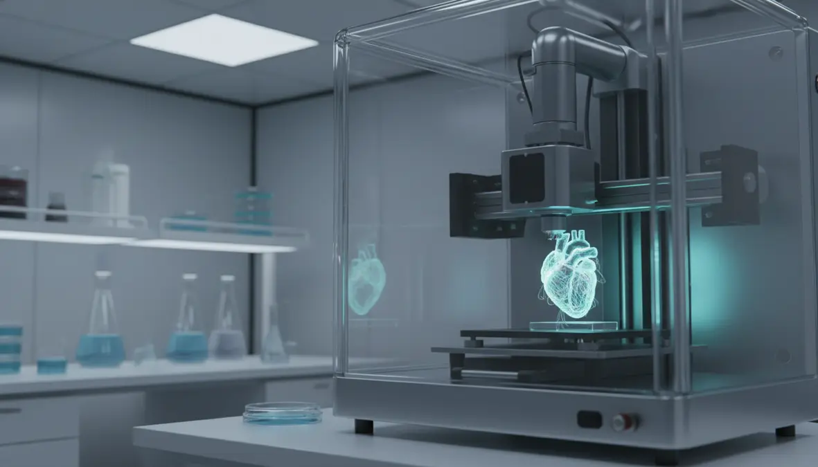
3D bioprinting is an additive manufacturing process that deposits living cells, biomaterials, and growth factors — collectively called bio-inks — layer by layer to create functional biological structures. Unlike traditional 3D printing that works with plastics or metals, bioprinters work with hydrogels loaded with millions of living cells, precisely positioning them in architectures that mimic natural tissue. The printer's nozzle extrudes bio-ink at micron-level accuracy, building structures that cells then colonize, remodel, and mature into living tissue over days or weeks in bioreactors.
Three core bioprinting technologies dominate in 2026. Extrusion-based bioprinting pushes bio-ink through a nozzle like a highly precise syringe, ideal for large tissue constructs. Droplet-based bioprinting (inkjet) deposits tiny droplets with cell-level precision, suited for complex multi-cell-type patterns. Light-based bioprinting — including digital light processing (DLP) and two-photon polymerization — uses focused light to solidify photosensitive bio-inks at resolutions below 10 micrometers, enabling the fabrication of capillary-scale vascular networks that were impossible just two years ago.
| Company | Technology | Key Achievement (2026) | Primary Application |
|---|---|---|---|
| Organovo | Extrusion bioprinting | FDA-cleared bioprinted liver tissue patches for chronic liver disease | Therapeutic implants |
| CELLINK (BICO) | Multi-material extrusion | BIO X 7 platform printing 6 cell types simultaneously | Research & drug testing |
| Aspect Biosystems | Microfluidic bioprinting | Bioprinted pancreatic tissue entering Phase II clinical trials for Type 1 diabetes | Organ therapeutics |
| Prellis Biologics | Holographic light-based | Printing vascularized tissue at 300 μm capillary resolution | Vascularized organs |
| CollPlant Biotechnologies | Plant-derived rhCollagen bio-ink | Bioprinted breast tissue implants in Phase I trials | Reconstructive surgery |
| Poietis | Laser-assisted bioprinting | Bioprinted skin models for L'Oréal cosmetics testing | Cosmetics & pharma |
The Vascularization Breakthrough
For years, the greatest barrier to bioprinting functional organs was vascularization — creating the intricate network of blood vessels needed to deliver oxygen and nutrients to cells deep within a tissue construct. Without vasculature, bioprinted tissue thicker than roughly 200 micrometers would die from the inside out. In 2026, that barrier has fallen.
Prellis Biologics' holographic bioprinting platform uses multiple laser beams to simultaneously polymerize bio-ink at thousands of points within a volume, building capillary networks at a speed and resolution that extrusion printers cannot match. Their latest published results demonstrate perfusable vascular trees with vessel diameters as small as 300 micrometers — approaching the scale of human arterioles. Researchers at Harvard's Wyss Institute have combined embedded bioprinting with sacrificial inks that dissolve after printing, leaving behind hollow channels that endothelial cells colonize to form functional blood vessels. The result: tissue constructs over 1 centimeter thick that remain viable for months.
Skin & Wound Healing
Bioprinted skin grafts using patient-derived keratinocytes and fibroblasts are now in clinical use for severe burns and chronic wounds. These grafts integrate faster than cadaver skin, produce natural pigmentation, and eliminate immune rejection entirely.
Cartilage & Bone
Bioprinted cartilage implants for knee and ear reconstruction have completed Phase III trials in Australia and South Korea. Using chondrocyte-laden hydrogels reinforced with biodegradable polymers, these implants gradually integrate with native cartilage over 6-12 months.
Organ-on-a-Chip
Bioprinted multi-organ chips connecting miniature liver, kidney, heart, and lung tissues are replacing animal testing in pharmaceutical R&D, with Emulate and CN Bio delivering FDA-accepted platforms that predict human drug toxicity with over 85% accuracy.
Transforming Drug Discovery
Perhaps the most immediate commercial impact of 3D bioprinting is in pharmaceutical research. Drug development is notoriously expensive — bringing a single drug to market costs an average of $2.6 billion and takes over a decade, with a 90 percent failure rate in clinical trials. A major reason for failure: preclinical models based on animal testing and 2D cell cultures poorly predict how drugs will behave in human tissue.
Bioprinted human tissue models change this equation fundamentally. Pharmaceutical giants including Roche, Johnson & Johnson, AstraZeneca, and Merck now use bioprinted liver, kidney, and tumor models for early-stage drug screening. These 3D tissue constructs replicate human physiology — cell-cell interactions, extracellular matrix composition, mechanical properties, and metabolic pathways — with a fidelity that 2D cultures and animal models cannot match. In January 2026, AstraZeneca published results showing that bioprinted tumor models predicted clinical trial outcomes for three oncology drugs with 87 percent accuracy, compared to 42 percent for traditional xenograft mouse models.
"Bioprinted tissue models are not incremental improvements over existing preclinical tools — they are a paradigm shift. We are seeing drug candidates fail or succeed in the dish the same way they fail or succeed in patients. That knowledge, obtained months or years earlier, is worth billions."
AI Meets Bioprinting
Artificial intelligence is accelerating bioprinting capabilities dramatically. Machine learning models trained on thousands of print runs now optimize bio-ink formulations, predict cell viability under different printing parameters, and design scaffold architectures that maximize nutrient diffusion and mechanical strength. Computer vision systems monitor the printing process in real time, detecting cell damage, nozzle clogging, and structural defects and adjusting parameters mid-print.
Generative AI is designing tissue architectures that no human engineer would conceive. DeepMind's collaboration with University College London uses neural networks to generate vascular network geometries optimized for oxygen delivery, producing branching patterns that outperform hand-designed architectures by 35 percent in perfusion efficiency. NVIDIA's BioNeMo platform provides foundation models for molecular biology that help researchers design novel bio-ink components — synthetic peptides, crosslinking agents, and growth factor release profiles — computationally before synthesizing them in the lab.
The Road to Whole Organ Printing
The ultimate goal of bioprinting — fabricating full transplantable organs like kidneys, livers, and hearts — remains years away but is closer than ever. The kidney is the most likely first candidate: its repetitive nephron architecture lends itself to scalable bioprinting approaches, and kidney disease affects over 850 million people globally, with transplant waitlists averaging 3-5 years in most countries. United Therapeutics, in partnership with 3D Systems, has invested over $300 million in a bioprinted organ program targeting a functional kidney prototype by 2028.
The challenges remaining are significant but tractable: scaling print volumes from cubic centimeters to the hundreds of cubic centimeters needed for a full organ, achieving the cellular density of native tissue (roughly 100 million cells per cubic centimeter for liver), and ensuring long-term functional maturation. Bioreactor technology — where printed constructs are cultured under physiological conditions including pulsatile flow, mechanical loading, and biochemical stimulation — is advancing rapidly, with constructs now maturing for 60-90 days while maintaining viability and developing tissue-specific function.
Market Outlook and What Comes Next
The global 3D bioprinting market, valued at $2.1 billion in 2025, is projected to reach $7.8 billion by 2030 according to Grand View Research, driven by pharmaceutical adoption, clinical translation of tissue products, and declining hardware costs. Bioprinter prices have fallen from over $200,000 in 2020 to under $40,000 for research-grade systems in 2026, democratizing access for universities and biotech startups worldwide.
- 2026-2027: FDA clearance of additional bioprinted tissue products (corneal implants, nerve guides, dental bone grafts). Widespread adoption of bioprinted tissue models in pharmaceutical R&D.
- 2028-2029: First bioprinted kidney tissue patches for partial organ repair enter clinical trials. In-situ bioprinting — printing tissue directly onto wounds during surgery — becomes standard for burn units.
- 2030 and beyond: Prototype whole-organ bioprinting for transplantation. Personalized medicine using patient-specific bioprinted tissue for treatment planning.
3D bioprinting is no longer a laboratory curiosity. It is a clinical reality for skin and cartilage, a commercial revolution for drug testing, and an accelerating pathway toward solving the global organ shortage that claims a life every eight minutes. The convergence of advanced bio-inks, AI-driven design, light-based printing, and vascularization breakthroughs means that the question is no longer whether we will print human organs — but when. And in 2026, that timeline is shorter than anyone expected.